Biological segmentation in WormAtlas
using machine learning segmentation methods to separate cells in C.elegans, quantifying segmentation uncertainty
C.elegans are an important “model organism” in biology and neuroscience as they were one of the first creatures to have had their full neuron structure mapped (e.g. their connectome)
As part of an NIH funded grant, the team is working with the WormAtlas Group on methods to segement cells and neurons in high resolution slices of these worms called Electron Microscopy (EM) Images.

While most segmentation methods assume a color image (e.g. 3-channel red, green blue), most EM images are a single gray-scale color channel.
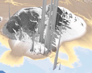
Luckily, we have a prior model that is up to this task – CloudFindr which was trained on digitial elevation maps taken from a satellite flying over the Arctic.

Work on the C.elegans version of our Cloudfindr model is ongoing – check back here soon for more details and updates!
References
2021
-
 CloudFindr: A Deep Learning Cloud Artifact Masker for Satellite DEM DataIn 2021 IEEE Visualization Conference (VIS) , Jan 2021
